
Supernumerary marker chromosomes – what they are, frequency and implications in newborns
25/11/2021
Share it:
![]()
![]()
![]()
![]()
![]()
Posted by:
Dr.M.Raszek
Chromosomal diversity – going beyond the 46
Recently, Merogenomics was referred to a person with a past medical history of unfortunate complications in a child and wanted to obtain second opinion on their unusual karyotype finding that analyzed that person’s chromosomes. That individual wanted to learn if these results could have a bearing on future attempts of having healthy children.
Karyotyping is the famous procedure of isolating and staining entire chromosomes which allows broad analysis of potential chromosomal abnormalities in an individual. It is a standard in the world of genetics (when genetic information is analyzed visually on this broad scale for potential abnormalities, it is referred to as cytogenetics) because it has been in use for a very long time. It is not a sensitive procedure though, meaning it can only detect structural changes to the chromosomes up to certain size because below that size, the changes are simply beyond visual confirmation. We cannot see it, and we need a better “microscope”. But it is still frequently used since it is such a well characterized procedure.
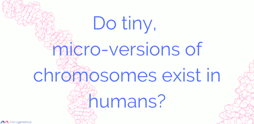
What was unusual about this finding was that it indicated the presence of what is referred to as supernumerary marker chromosome or the addition of an independent fragment of a chromosome derived from any of our naturally occurring chromosomes. In essence, a person ends up having an additional tiny chromosome, and the clinical consequences of this can be quite varied. More about that in a moment. Normally human genomes are made up of 46 chromosomes. Each chromosome is present in pairs, apart from the sex chromosomes in males, who present with unique X and Y chromosomes (whereas females will have two X chromosomes). Another way to say this, is that there are 24 different human chromosomes: 22 chromosomes plus X and Y sex chromosomes. We inherit 22 of these chromosomes plus one sex chromosome from each of our parent’s gametes, to produce a total of 46 chromosomes as part of our genomes. Thus a regular karyotype would observe these 46 chromosomes (labelled as 46,XX to indicate a female or 46,XY to indicate a male).

This regular pattern of chromosomes can frequently be imbalanced, either by a loss or addition of an entire chromosome (collectively referred to as aneuploidy, where a chromosome loss is referred to as monosomy, and a chromosomal gain is referred to as trisomy). Another event that can take place is a duplication or loss of the fragment of a chromosome within an existing chromosome, thus leading to imbalance but without affecting the number of chromosomes. A segment of a chromosome can also be inverted in place, where its normal direction is reversed (called inversion). You can also have fragments of chromosomes swapping places between different chromosomes. For example, chromosome 9 and chromosome 22 can break and exchange portions, an event that predisposes an individual to chronic myelogenous leukemia, the most famous example of acquired chromosomal change leading to malignancy. This particular event is referred to as the Philadelphia chromosome, named after the city where it was first described. Finally, you can also accidentally have one extra version of every chromosome, and that is referred to as triploidy. In essence, an individual has an entire extra set of chromosomes for a total of 69 chromosomes. This can happen for example if an egg is accidentally fertilized by two sperm cells. Even tetraploidies are possible (four sets of all chromosomes for a total of 92 chromosomes!). Human genomes can be extremely diverse although typically the disruption of chromosomal balance frequently results in clinical consequences.
Extra tiny chromosomes
However, in addition to the above well characterized events, there is also a possibility of an accidental generation of a small fragment of any one of the 24 different human chromosomes, leading to the formation of supernumerary marker chromosome. Surprisingly, these events apparently are quite frequent, so it was a weird to us that we did not really hear about their detection previously in all the literature we studied in relation to non-invasive prenatal testing (NIPT). Apparently supernumerary marker chromosomes are observed in 1:1,340 prenatal examinations and in 1:2,250 newborns, which is very frequent and not that different from the frequency of Down syndrome. One of the authors of that publication, Dr. Thomas Liehr (who is available for a consult), compiles information on these events and has formed a privately generated database on marker chromosomes. This database is a very valuable resource for genetic counselors and medical geneticists attempting to discern potential outcomes of different marker chromosomes. Marker chromosomes are also a contributing factor to infertility, especially in men, with 0.125% infertile individuals presenting with these unusual events (nearly a 3-fold increased risk over the general population), with men outnumbering women to the tune of 7.5-fold. Age is a contributing factor to incidence rate. The rate is even higher in patients with intellectual disability, accounting for 0.288% of such cases.
Below is a visual of a karyotype as observed in the Merogenomics referral (presented with permission).

In this privately obtained karyotype you can clearly see 46 chromosomes with X and Y chromosomes indicated male gender, and a presence of a marker chromosome (such karyotype would be abbreviated as 47,XY+MAR with 47 indicating total number of chromosomes found; XY being part of those 47 chromosomes; and +information presenting what additional chromosomal content has been discovered. Ideally, the chromosomal origin of the marker chromosome could be provided because such information could then be attempted to be correlated to potential clinical outcomes. The unfortunate part is that the treating physician of this individual did not understand these results and assumed they were incorrect, thinking they were supposed to indicate the presence of Down syndrome (produced most frequently by a trisomy of chromosome 21 and on rare occasions, by duplication of one segment of chromosomes 21) whereas the individual with this karyotype was clinically normal. The complications observed in a child were not linked by the doctor to the potential significance of this finding. Instead, the family suspected that perhaps there are other genetic predispositions for the clinical outcomes observed in a child and wanted to undergo genomic testing. Merogenomics sounded the alarm for this person, and recommended seeking genetic counseling because marker chromosomes can lead to negative outcomes. Genome sequencing could identify which chromosome might have been the source of the marker chromosome, but such information could also be gained with a chromosomal microarray and FISH (also part of the cytogenetics family of tests) that could be called on as part of standard healthcare. FISH is fluorescence in situ hybridization, where you light up a specific area of a chromosome with a fluorescent dye and then look under microscope. You can inspect multiple chromosomes with different colored dyes and catch unusual chromosomal events. These procedures can analyse information on an even deeper level than a karyotype, hence acting like that “microscope” mentioned earlier. We wanted this individual to first discover what options were available to the family as part of the public healthcare.
What are the implications of marker chromosomes?
Despite the apparent frequency of marker chromosomes, their impact is still quite mysterious, and very difficult to predict an outcome. This is because marker chromosomes are still understudied, despite their apparent high rate of occurrence. Genome sequencing technologies are bound to rapidly advance that knowledge, though. According to Dr. Liehr’s information, about 70% of the cases with marker chromosomes are de novo (meaning they originated spontaneously in the gametes, either sperm or egg), and the remaining 30% are inherited from one of the parents. About 30% of the carriers of marker chromosomes are going to present with clinical complications, while 70% are expected to be clinically normal.
As with anything related to DNA, how these marker chromosomes arise is complex and many different types of marker chromosomes can be produced, including unusual circular chromosomes. Other common possibilities include microscopic chromosomes (tiny chromosomal fragments) and another unusual formation, called inverted, duplicated chromosomes. In this case, imagine a tiny small chromosome that itself is duplicated with both of these chromosomes attached together by the same end, as if the duplicated fragments bound together were mirroring each other.
One method of how marker chromosomes are produced appears to be due to a rescue attempt if fertilization took place with a gamete that accidentally had an extra chromosome (trisomy). Trisomy happens when during gamete production, the chromosomes are not properly divided equally between the daughter cells, leading one of the gametes to carry two chromosomes instead of one. During fertilization one gamete provides one chromosome, and the other gamete provides two chromosomes instead of one, leading to trisomy. Sometimes such accidental trisomies are attempted to be rescued in the zygote by having the extra chromosome destroyed. Incomplete destruction of such trisomic chromosomes can lead to a remaining marker chromosome. This appears to be especially common in de novo marker chromosomes. Without this attempted rescue from trisomy, the trisomic fetus would have high likelihood of not surviving.

While every single chromosome can be affected, most marker chromosomes originate from acrocentric chromosomes, or chromosomes that have their centromeres positioned near the end of the chromosome. Centromeres are a special area of DNA that is used to split duplicated chromosomes apart during cell division, when a new cell is being produced from a parent cell. The estimate is that 70% of marker chromosomes derive from these type of chromosomes. 55-59% of marker chromosomes are also present in a mosaic state. Mosaicism describes the existence of different unique genomes making up a single individual. In this case it would mean that the marker chromosome is only present in a subset of tissues making up that person.
The frequency of these events actually really surprised us because we have not encountered these described in the literature dedicated to non-invasive prenatal testing (NIPT) but this might then account for some of the assumed test failures. NIPT is the most powerful “microscope” technology, if you will. NIPT looks at amounts of fragmented DNA circulating in the blood to count any imbalances in chromosomes of the placenta (which indirectly informs us of the chromosomal state of the fetus). But as mentioned, marker chromosomes can occur from any chromosome, and this may be the underappreciated power of using a full NIPT (also called expanded NIPT, when all chromosomes are investigated at the same time, rather than just the most three common trisomies: 13, 18 and 21). NIPT has the capacity to provide detailed information on chromosomal abnormalities, including marker chromosomes. A potential problem might be that NIPT could inaccurately point to a trisomy as opposed to marker chromosome that is mosaic, but it is for this reason that NIPT positive results are always confirmed with invasive test.
As authors of one rare publication on NIPT with marker chromosome noted, when NIPT results suggestive of marker chromosome are returned, “it is extremely important to perform not only NIPT verification on a sample of material other than trophoblast, but also to apply appropriate research methods.” Thus, it is important to confirm NIPT with a different source of genetic content (from a different type of cells) and to smartly select your tools of analysis to accurately confirm the result. FISH alone might not be sufficient for a diagnosis, requiring additional tools. But also, the choice of cells selected for subsequent confirmatory tests may be important! In this case the authors described a case where placental cells that were uncultured versus cultured provided different results. In uncultured cells from the invasive sample the presence of normal cells was indicated, also cells with trisomy and finally cells with a marker chromosome originating from same chromosome that was causing trisomy in the other cells. The cultured cells, on the other hand, only showed normal cells or cells with a marker chromosome only. Trisomic cells disappeared and the authors thought this difference was due to that same trisomy rescue where cells had the opportunity to grow during culturing and that led to the emergence of a marker chromosome early in the development. It shows you how much nature wants to fight to restore the chromosomal balance (for increased survival chances).
In conclusion, marker chromosomes are unique but not rare events with a significant likelihood of clinical consequences to the developing fetus. After discovery, very careful analysis of any marker chromosomes should be ongoing to help facilitate better risk analysis towards the clinical outcomes of such events. A full, expanded NIPT might be instrumental in identifying such events, especially if ultrasounds are uninformative of any developing problems.
Merogenomics would like to thank their client for permission to bring to light this overlooked condition of marker chromosomes and we will continue to do our best to find research that correlates presentations with outcomes and also especially to continue to inform and present to doctors and patients by writing blogs, giving online help sessions, and educating on the merits of clinical DNA testing.
This article has been produced by Merogenomics Inc. and edited by Jason Chouinard, B.Sc. Reproduction and reuse of any portion of this content requires Merogenomics Inc. permission and source acknowledgment. It is your responsibility to obtain additional permissions from the third party owners that might be cited by Merogenomics Inc. Merogenomics Inc. disclaims any responsibility for any use you make of content owned by third parties without their permission.
Products and Services Promoted by Merogenomics Inc.
Select target group for DNA testing
Healthy screening |
Undiagnosed diseases |
Cancer |
Prenatal |
Or select popular DNA test
 |
 |
 |
 |
Pharmaco-genetic gene panel |
Non-invasive prenatal screening |
Cancer predisposition gene panel |
Full genome |

Clinical DNA testing
genome sequencing consulting
www.merogenomics.ca
info [at] merogenomics [dot] ca
Edmonton, Canada
Disclaimer:
All information presented is for educational purposes only (including provided links). No information presented by Merogenomics Inc. should be used in place of advice or consultation with a healthcare provider. NEVER DISREGARD PROFESSIONAL MEDICAL ADVICE ON ACCOUNT OF INFORMATION PROVIDED BY MEROGENOMICS INC.
Merogenomics Inc. does not guarantee the efficacy of the products or services it mentions (whether endorsed or not), or the accuracy, completeness, relevance, and novelty of all presented information, and makes no representation or warranty to such guarantees. Information presented is subject to change at any time without notice to reflect novel concepts or improvement to offered services. The material presented by Merogenomics Inc. about the utility of genome sequencing technologies can be cutting-edge and forward-looking, and not acceptable to everyone. You agree and acknowledge that your study and utility of information presented by Merogenomics Inc. is undertaken at your own risk.
This site contains images generated by emovie combined with pymol software.